Retinal detachment
Disease description
Retinal detachment is an ophthalmological pathology in which the outer layers of the retina, containing rods and cones responsible for color perception, separate from the deeper layers. This condition is serious and requires immediate medical attention.
Retinal detachment occurs more frequently in individuals with myopia. Patients with diabetes mellitus, arterial hypertension, and eye injuries are also at risk.
Symptoms indicating the need for diagnosis and treatment
The following symptoms suggest retinal detachment:
- A veil over the eyes (which does not disappear when rubbing the eye)
- Flashes of light in the eye (electrical impulses at the peak of detachment)
- Loss of visual field areas
- Blurred vision and unclear contours
Patients often report that their vision appears sharper upon waking up in the morning than during the day. This occurs because the retinal layers are touching each other in a horizontal position.
Diagnostic and treatment methods
Diagnostic
According to modern clinical guidelines used in top European clinics, patients suspected of retinal detachment undergo:
- Visometry and perimetry (to assess visual acuity and field of vision)
- Tonometry (to measure intraocular pressure)
- Ophthalmoscopy (to examine the fundus of the eye)
- Ultrasound and Optical Coherence Tomography (OCT)
Treatment
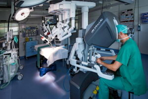
Retinal detachment always requires urgent microsurgery, which is performed using the following methods: local sealing of the tear (for partial detachment), encircling scleral buckling (for complete detachment), vitrectomy with subsequent replacement of the vitreous body and laser therapy.
Innovations in global clinics
The AI is now used in retinal image segmentation alongside tomography — a unique method that allows for quick detection of detachment and informs the doctor via a special encoded signal. Once flagged, the ophthalmologist reviews the scan again to confirm the diagnosis.
Procedures
Top clinics
-
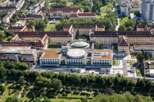

 University Hospital Freiburg
 -

 Acibadem Altunizade Clinic
 -

 Ajibadem Atasehir Clinic
 -
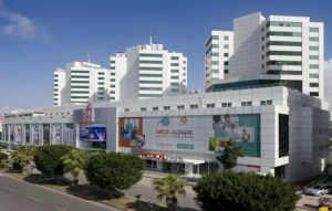

 Medical Park Antalya Clinic
 -

 Dubai, UAE NMC Healthcare
 -

 Shishli Memorial Clinic.
 -
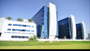 Al Zahra Hospital
 -

 Milan, Italy San Raffaele University Hospital
 -
 American Hospital Dubai
 -
 Leech Private Clinic
 -
 Burjeel Hospital Abu Dhabi
 -

 Debling Private Clinic
 -
 Confraternity Private Clinic.
 -
 Burjeel Hospital Private Multidisciplinary Clinic
 -
 University Hospital Heidelberg
 -

 Vienna, Austria Wiener Privatklinik (WPK)
 -

 Oberhausen Clinic of the Niederrein Complex
 -
 Munich, Germany University Hospital Munich (Ludwig-Maximilians-Universität)
 -

 Charité Clinic
 -

 Bahçelievler Memorial Clinic
 -
 Hangil Ophthalmology Clinic
 -
 Clinique Montchoisy
 -
 Clinique Genolier
 -

 Ataşehir Memorial Clinic
 -

 Memorial Antalya Hastanesi
 -
 Acibadem Bodrum Hospital
 -
 Zurich, Switzerland Bethanien Clinic
 -

 Barcelona, Spain QuironSalud Barcelona Hospital
 -
 Barcelona, Spain Dexeus University Hospital
 -
 Barcelona, Spain Medical Center "Teknon"
 -
 Barcelona, Spain Sant Joan de Deu Children's Hospital
 -
 Barcelona, Spain University Hospital Barnaclinic+
 -

 Madrid, Spain University Clinic HM Madrid
 -
 Madrid, Spain University Hospital HM Monteprincipe
 -
 Düsseldorf, Germany Breyer, Kaymak, and Klabe Eye Surgery Clinic
 -
 Hamburg, Germany Asklepios Klinik Barmbek
 -
 Gebze, Turkey Anadolu Clinic
 -
 Zurich, Switzerland Hirslanden Clinic
 -
 Madrid, Spain Quiron Salud University Hospital
 -

 Seoul, South Korea Samsung Medical Center
 -
 Bursa, Turkey Doruk Nilüfer Hospital
 -
 SNUH
SNUH -

 г. Женева, Швейцария Клиника «Женераль-Болье»
 -
 г. Женева, Швейцария Hirslanden Clinique La Colline
 -
 г. Стамбул, Турция Клиника Флоренс Найтингейл
 -
 г. Сеул, Южная Корея Медицинский центр «Асан»
 -

 г. Иерусалим, Израиль Медицинский центр “Хадасса”
 -
 г. Петах-Тиква, Израиль Медицинский центр имени Ицхака Рабина
 -
 г. Тель Авив, Израиль Медицинский центр “Ассута”
 -

 г. Рамат-Ган, Израиль Клиника Шиба