Melanoma
Melanoma is a malignant condition that arises from the pigment-producing skin cells — melanocytes — due to their abnormal transformation and uncontrolled proliferation. Melanoma is considered the most aggressive type of skin cancer. Early detection of the tumor can bring the five-year survival rate after treatment to approximately 98%.
Types of melanomas:
Superficial spreading melanoma. The most common form, accounting for about 70% of all cases. Typically diagnosed between the ages of 30 and 50, more often in women of Caucasian descent. Prognosis is favorable with early detection.
Nodular melanoma. The second most common type (up to 20% of cases). Usually arises on previously unaffected skin and is more prevalent in men aged 40-60. It grows vertically from the outset, infiltrating deep tissues. The prognosis is less favorable.
Lentiginous melanoma. Accounts for 5-10% of all melanoma cases. More commonly diagnosed in women at an older age (60-70 years). Appears as a pigmented macule and is less likely to metastasize than other types.
Amelanotic melanoma. The rarest type (1-2% of all melanomas). Presents as a small, rough nodule on the skin, often located on the fingers, soles, or heels. It lacks pigment and is therefore harder to detect visually.
Symptoms indicating the need for diagnosis and treatment
Melanomas most often develop from pre-existing nevi (moles) that undergo malignant transformation under the influence of carcinogenic factors such as heredity and ultraviolet radiation.
Cutaneous malignancies are not always visible to the naked eye, so any suspicious changes in moles require consultation with an oncologic dermatologist.
Warning signs in a nevus (mole) include:
- Asymmetry in shape
- Enlargement in diameter to over 6 mm
- Color changes (such as speckled or uneven pigmentation)
- Irregular or jagged borders
- Sensations of pain or itching
- Thickening, bleeding, or crusting of the surface
Diagnostic and treatment methods
Diagnosis
Currently, the clinical diagnostic accuracy of primary melanoma in advanced clinics reaches up to 98%. Diagnostic methods include: blood tests for specific tumor markers, squamous cell carcinoma antigen (SCC) testing, biopsy (histological examination of a tissue sample), Computed Tomography (CT), Magnetic Resonance Imaging (MRI).
Treatment
The primary approach to treating melanoma is radical surgical excision with wide margins of healthy tissue. If metastases are present, the following treatments are also employed:
- Polychemotherapy — particularly effective in stage III and higher
- Radiation therapy — commonly used postoperatively
- Immunotherapy — aimed at stimulating the patient’s own immune system
- Targeted therapy — a modern method for treating metastatic melanoma
Innovations in global clinics
Researchers from the Laboratory of Radiation Biology have developed a fundamentally new technique to enhance the effectiveness of radiation therapy by intensifying proton beams to suppress melanoma growth. This method targets tumor stem cells and reduces radioresistance.
Top clinics
-
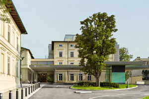
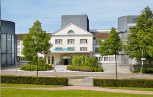
 Max Grundig Clinic
 -
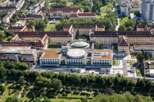


 University Hospital Freiburg
 -

 Acibadem Altunizade Clinic
 -
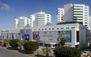

 Medical Park Antalya Clinic
 -
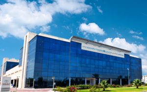

 Dubai, UAE NMC Healthcare
 -
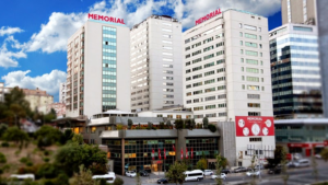
 Shishli Memorial Clinic.
 -
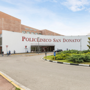

 Milan, Italy San Donato Hospital in Milan, Italy
 -
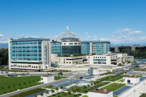 Milan, Italy San Raffaele University Hospital
 -
 American Hospital Dubai
 -
 Burjeel Hospital Abu Dhabi
 -

 Debling Private Clinic
 -
 Rudolfinerhaus Private Clinic.
 -
 University Hospital Heidelberg
 -

 Vienna, Austria Wiener Privatklinik (WPK)
 -
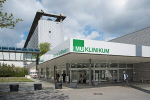 Munich, Germany University Hospital Munich (Ludwig-Maximilians-Universität)
 -

 Asklepios Nord Heidberg
 -
 FKKD Clinical Complex
 -

 Charité Clinic
 -

 Gil Medical Center at Gachon University
 -
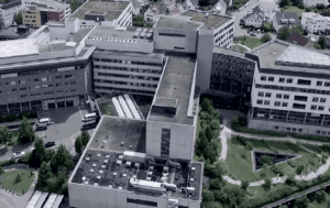
 Clinique Genolier
 -

 Barcelona, Spain QuironSalud Barcelona Hospital
 -
 Barcelona, Spain Medical Center "Teknon"
 -
 Barcelona, Spain University Hospital Barnaclinic+
 -

 Madrid, Spain University Clinic HM Madrid
 -
 Madrid, Spain University Hospital HM Monteprincipe
 -
 Hamburg, Germany Asklepios Klinik Barmbek
 -
 Gebze, Turkey Anadolu Clinic
 -
 Zurich, Switzerland Hirslanden Clinic
 -
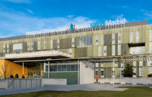 Madrid, Spain Quiron Salud University Hospital
 -
 Duesseldorf, Germany Oncological Center Dusseldorf
 -

 Seoul, South Korea Samsung Medical Center
 -
 Seoul, South Korea Medical Center at Ewha Womans University
 -
 SNUH
SNUH -

 г. Женева, Швейцария Клиника «Женераль-Болье»
 -
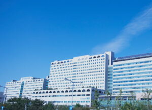
 г. Сеул, Южная Корея Медицинский центр «Асан»
 -

 г. Иерусалим, Израиль Медицинский центр “Хадасса”
 -
 г. Петах-Тиква, Израиль Медицинский центр имени Ицхака Рабина
 -
 г. Тель Авив, Израиль Медицинский центр “Ассута”
 -
 г. Рамат-Ган, Израиль Клиника Шиба