Rhabdomyosarcoma
Disease description
Rhabdomyosarcoma (RMS) is a rare and aggressive type of cancer that arises from skeletal muscle cells. Rhabdomyosarcoma primarily affects children under the age of 10, although it can also develop during adolescence.
RMS can occur in any part of the body where muscle tissue is present. The most common locations include:
- head and neck region: the tumor may develop near the eyes, in the paranasal sinuses, throat, or even in the ear region
- genitourinary tract: it can affect the urinary bladder, prostate gland, vagina, and uterus
- extremities: the malignancy may arise in the muscles of the arms and legs
- trunk: the tumor can develop in the chest wall, abdominal cavity, or pelvic region
Symptoms
Rhabdomyosarcoma is often detected at early stages, particularly because the tumor frequently develops in visible or palpable areas. In approximately one-third of cases, the tumor is diagnosed at a stage when it can be almost entirely removed surgically (though microscopic metastases or residual tumor tissue requiring further treatment with chemotherapy are often present). Fewer than 20% of patients present with distant metastases at the time of diagnosis. The symptoms of RMS largely depend on the location of the primary tumor:
- Subcutaneous tumor: appears as a visible swelling or is palpable as a firm mass beneath the skin.
- Orbital tumor: proptosis (bulging of the eye), strabismus (misalignment of the eyes), and possibly complaints of diplopia (double vision).
- Nasal cavity tumor: nasal congestion, epistaxis (nosebleeds), or bloody/mucoid nasal discharge.
- External auditory canal tumor: otorrhea (ear discharge) and/or hearing loss.
- Bladder tumor: dysuria (difficulty urinating), hematuria (blood in the urine).
- Genital tract tumor:
○ In boys: scrotal swelling.
○ In girls: bloody or mucous vaginal discharge. - Abdominal or pelvic tumor: abdominal pain, constipation, vomiting.
- Extremity tumors (often alveolar RMS in adolescents): may be mistaken for a contusion; therefore, any swelling that enlarges rapidly or fails to resolve over several weeks requires medical evaluation.
Diagnosis and treatment for rhabdomyosarcoma in children
Diagnostics
If rhabdomyosarcoma is suspected, the following diagnostic procedures are performed:
- Biopsy: necessary to obtain a tissue sample for histopathological examination and to determine the RMS subtype.
- Histopathological examination: microscopic analysis of the tumor tissue to identify specific cellular features and genetic mutations associated with RMS.
- Immunohistochemical analysis: performed to detect cellular markers characteristic of rhabdomyosarcoma.
- Cytogenetic and molecular genetic studies: used to confirm the diagnosis and identify chromosomal abnormalities such as translocations t(1;13) or t(2;13), which are typical of the alveolar subtype.
- Imaging studies:
- Ultrasound, CT scan, and MRI: employed to assess the size of the tumor, its invasion into surrounding tissues, and presence of metastases.
- Chest radiography: conducted to detect lung metastases, one of the common sites of dissemination in RMS.
- Bone scintigraphy: used to identify skeletal metastases.
- Bone marrow analysis: performed through bone marrow aspiration and biopsy to detect or exclude metastases to the bone marrow.
- Lumbar puncture: indicated if the tumor is located close to the meninges of the brain or spinal cord, to evaluate cerebrospinal fluid for malignant cells.
Treatment
The treatment strategy for rhabdomyosarcoma is determined by multiple factors, including disease stage, tumor histological subtype, patient age, and tumor location. Treatment typically consists of surgery and chemotherapy, and in most cases, radiation therapy is also employed.
For tumors located in the head and neck region, surgical intervention may be complicated due to proximity to vital structures. This requires an individualized surgical approach, as wide excision of surrounding healthy tissue may not always be feasible. Such cases may necessitate the involvement of neurosurgeons and specialists in vascular or reconstructive surgery.
Neoadjuvant chemotherapy (administered prior to surgery) is often used to shrink the tumor and facilitate surgical resection. Adjuvant chemotherapy is mandatory after surgery — even if the tumor is completely removed — to eradicate residual malignant cells and minimize the risk of recurrence. In some cases, maintenance chemotherapy may be prescribed following the primary treatment course.
Radiation therapy is generally used to destroy remaining cancer cells after surgery and chemotherapy, but it may also be initiated prior to surgery in certain cases. When the tumor is located near the meninges of the brain or spinal cord, radiation therapy may begin immediately after diagnosis. Specialized radiation modalities, such as proton therapy and brachytherapy, may be recommended depending on the tumor’s location and characteristics.
Innovations in global clinics
In 2022, researchers achieved a breakthrough in the treatment of rhabdomyosarcoma by developing a targeted drug — an inhibitor of the enzyme KDM4B, which is responsible for promoting aggressive tumor growth.
In clinical trials, the combined use of the KDM4B inhibitor with standard chemotherapy led to complete tumor regression in several cases. This targeted agent is now being used for the treatment of complex and chemotherapy-resistant forms of rhabdomyosarcoma in children and teens in Europe and the United States.
Top clinics
-


 Baden-Baden, Germany Max Grundig Clinic
 -

 Seoul, South Korea Asan Medical Center
 -

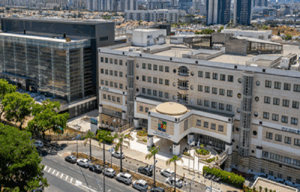
 Jerusalem, Israel Hadassah Medical Center
 -
 Petah Tikva, Israel Medical Center “Rabin”
 -
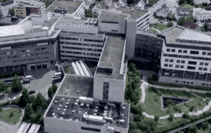
 Geneva, Switzerland Hirslanden Clinique La Colline
 -

 Geneva, Switzerland Generale-Beaulieu
 -

 Istanbul, Turkey Acibadem Altunizade
 -
 Antalya, Turkey Hospital Medical Park Antalya
 -

 Dubai, UAE NMC Healthcare
 -
 Istanbul, Turkey Hospital “Memorial Şişli”
 -

 Milan, Italy San Raffaele University Hospital
 -
 Abu Dhabi, UAE Burjeel Hospital Abu Dhabi
 -


 Vienna, Austria Debling Private Clinic
 -
 Heidelberg, Germany Heidelberg University Hospital
 -
 Winterthur, Switzerland Clinic "Lindberg"
 -
 Nyon, Switzerland Clinique Genolier
 -

 Barcelona, Spain QuironSalud Barcelona Hospital
 -
 Barcelona, Spain Medical Center "Teknon"
 -
 Barcelona, Spain Sant Joan de Deu Children's Hospital
 -
 Barcelona, Spain University Hospital Barnaclinic+
 -

 Madrid, Spain University Clinic HM Madrid
 -
 Madrid, Spain University Hospital HM Monteprincipe
 -
 Zurich, Switzerland Hirslanden Clinic
 -
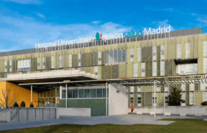 Madrid, Spain Quiron Salud University Hospital
 -
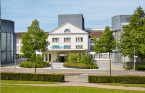
 Duesseldorf, Germany Oncological Center Dusseldorf
 -
 Petah Tikva, Israel Schneider Children's Medical Center
 -
 Seoul, South Korea Samsung Medical Center
 -
 SNUH
SNUH