

Skeletal scintigraphy
Skeletal scintigraphy is an important diagnostic method used in oncology to assess the condition of the skeletal system and detect metastases. This method enables physicians to identify cancerous lesions in the bones and evaluate the extent of the disease, which plays a key role in determining the treatment strategy.
Method description and its effectiveness
Skeletal scintigraphy, or bone scintigraphy, is a nuclear medicine imaging technique. During the procedure, the patient is injected with a small amount of a radioactive substance called a radiopharmaceutical. The radiopharmaceutical circulates through the bloodstream and accumulates in the bone tissue, particularly in areas of active bone metabolism, allowing the detection of potential metastases.
After the injection, the patient lies on a table, and a special gamma camera detects the radiation emitted by the radiopharmaceutical. The resulting images allow visualization of the bone condition and the identification of abnormal areas that indicate cancerous involvement.
Skeletal scintigraphy has a high sensitivity (approximately 80-90%) and specificity (about 70-80%) in detecting bone metastases. In some cases, especially when dealing with small lesions, the sensitivity may be lower. However, thanks to the use of advanced technologies in modern clinics, such as SPECT/CT, the diagnostic accuracy is significantly improved.
Advantages
- Preparation: before starting treatment, a detailed examination is carried out to determine the optimal chemotherapy regimen and select the appropriate drugs.
- Drug administration: chemotherapy can be administered in various ways:
○ Intravenously: drugs are delivered via an IV drip.
○ Orally: in the form of tablets or capsules.
○ Locally: drugs can be injected directly into the tumor or a body cavity. - Treatment course: chemotherapy is administered in cycles alternating with rest periods, allowing the body to recover. The number of cycles depends on the specific case and may vary.
- Monitoring and support: throughout the course of treatment, doctors closely monitor the patient’s condition to adjust dosages promptly and provide supportive care.
Features of the procedure in advanced clinics
In leading medical centers across Europe and worldwide, skeletal scintigraphy is performed using advanced gamma cameras that provide high-resolution images. Some clinics also employ hybrid methods such as SPECT/CT (Single Photon Emission Computed Tomography combined with Computed Tomography), which not only detect pathological changes but also precisely localize them in the body.
Moreover, the clinics place great emphasis on patient comfort. The procedure is carried out in a calm and relaxing environment, which helps reduce patient stress and anxiety.
Top clinics
-
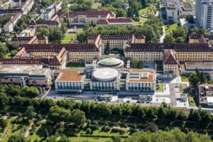

 University Hospital Freiburg
 -

 Acibadem Altunizade Clinic
 -
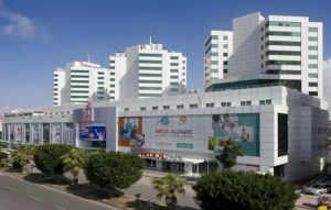

 Medical Park Antalya Clinic
 -
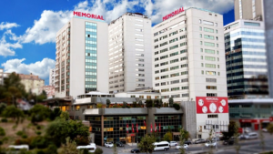
 Shishli Memorial Clinic.
 -
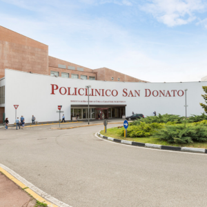

 Milan, Italy San Donato Hospital in Milan, Italy
 -
 Milan, Italy San Raffaele University Hospital
 -
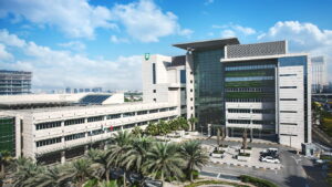 American Hospital Dubai
 -

 Debling Private Clinic
 -
 Confraternity Private Clinic.
 -
 Rudolfinerhaus Private Clinic.
 -

 University Hospital Heidelberg
 -

 Vienna, Austria Wiener Privatklinik (WPK)
 -

 Asklepios Nord Heidberg
 -
 Clinique Genolier
 -

 Barcelona, Spain QuironSalud Barcelona Hospital
 -
 Barcelona, Spain Dexeus University Hospital
 -
 Barcelona, Spain Medical Center "Teknon"
 -
 Barcelona, Spain Sant Joan de Deu Children's Hospital
 -
 Barcelona, Spain University Hospital Barnaclinic+
 -

 Madrid, Spain University Clinic HM Madrid
 -
 Madrid, Spain University Hospital HM Monteprincipe
 -
 Gebze, Turkey Anadolu Clinic
 -
 Zurich, Switzerland Hirslanden Clinic
 -
 Madrid, Spain Quiron Salud University Hospital
 -

 Seoul, South Korea Samsung Medical Center
 -
 SNUH
SNUH -

 г. Рамат-Ган, Израиль Клиника Шиба
 -
 г. Тель Авив, Израиль Медицинский центр “Ассута”
 -
 г. Петах-Тиква, Израиль Медицинский центр имени Ицхака Рабина
 -
 г. Иерусалим, Израиль Медицинский центр “Хадасса”