Neuroblastoma
Disease description
Neuroblastoma is an aggressive and rare form of cancer that develops in children, most commonly under the age of 5. This malignant tumor originates from immature nerve cells called neuroblasts, which are part of the sympathetic nervous system. This system controls the body’s response to stress, including increased heart rate, elevated blood pressure, pupil dilation, and other physiological changes.
Neuroblastoma most frequently develops in the adrenal glands — endocrine glands located above the kidneys that are responsible for producing stress hormones such as adrenaline and cortisol. However, the primary tumor may also form anywhere along the spine, for example, in the chest cavity (affecting the mediastinum, the central compartment of the thoracic cavity) or in the neck region.
Neuroblastoma has several distinctive features. It often presents as an aggressive malignancy capable of rapid growth and metastasis. However, in some cases — particularly in very young children — it may exhibit the opposite behavior: spontaneous regression, where the tumor gradually disappears without treatment. Additionally, neuroblastoma cells may sometimes “mature” (differentiate), transforming a malignant tumor into a benign one known as a ganglioneuroma.
Symptoms
The clinical presentation of neuroblastoma varies depending on the tumor’s location. If the tumor is located in the retroperitoneal space, abdominal swelling may occur, along with pain or discomfort. On palpation, a dense, nearly immobile mass may be detected.
A tumor in the thoracic cavity can cause coughing, difficulty breathing, trouble swallowing, and chest wall deformities. When the tumor extends into the spinal canal, it may lead to weakness or numbness in the legs, partial paralysis, as well as bladder and bowel dysfunction. If the tumor grows in the retrobulbar space (behind the eyeball), it may cause exophthalmos (eye protrusion) and periorbital bruising.
Tumor compression of blood and lymphatic vessels can lead to edema. In some cases, involuntary eye movements (opsoclonus) and muscle twitching (myoclonus) may occur.
Other general symptoms, common to many oncological diseases, may also be present: fever, elevated blood pressure, tachycardia, weight loss, loss of appetite, and sometimes diarrhea.
Neuroblastoma most commonly metastasizes to the bones, bone marrow, and lymph nodes. Bone metastases cause pain and limping. Bone marrow involvement results in a deficiency of blood cells, leading to anemia (paleness, fatigue), thrombocytopenia (increased bleeding), and leukopenia (weakened immune system). Lymphadenopathy (enlarged lymph nodes) may indicate metastasis. Cutaneous metastases may manifest as bluish or reddish lesions. Hepatic involvement leads to hepatomegaly (enlarged liver). Rarely, metastases may spread to the brain or lungs.
Diagnosis and treatment of neuroblastoma in children
Diagnosis
- Urine test for catecholamines
One of the key diagnostic tools for neuroblastoma is a urine test for catecholamines (epinephrine, norepinephrine, and dopamine). In the body, catecholamines are metabolized into homovanillic acid (HVA) and vanillylmandelic acid (VMA). In most cases of neuroblastoma, levels of HVA, VMA, and dopamine in the urine are significantly elevated, serving as biomarkers for the disease.
- Biochemical tests: Neuron-Specific Enolase (NSE)
In addition to urine tests, blood tests are conducted to determine the level of the enzyme neuron-specific enolase (NSE). NSE is a tumor marker used to assess tumor activity. Elevated levels of NSE may indicate the presence of neuroblastoma and its degree of aggressiveness.
- Imaging techniques: CT, MRI, and Scintigraphy
To precisely identify the tumor’s location and size, imaging techniques such as computed tomography (CT) and magnetic resonance imaging (MRI) are used. CT scans provide detailed images of internal organs and reveal tumor size and spread. MRI offers superior visualization of soft tissues and is particularly valuable in assessing the spinal cord and brain.
Scintigraphy using radionuclides is crucial for detecting bone metastases. This method involves the injection of a small amount of radioactive material, which accumulates in tumor cells and metastases, making them visible on a special scanner.
- Biopsy
To confirm the diagnosis, a biopsy is necessary — this procedure involves extracting a tissue sample from the tumor for histopathological examination. Biopsy identifies the tumor cell type and malignancy grade, making it a critical step in determining the most appropriate treatment plan.
- Molecular genetic analysis of tumor cells
Molecular genetic studies play a vital role in diagnosing and planning treatment. These include tests for the amplification and expression of the N-MYC oncogene. Identifying specific mutations helps predict tumor behavior and responsiveness to various therapies, allowing for a personalized treatment approach and improved outcomes.
Treatment
Treatment of neuroblastoma in children is a complex, multi-stage process involving both conventional and innovative methods. Leading global clinics use comprehensive treatment protocols, including chemotherapy, surgical intervention, radiotherapy, and targeted therapies.
Chemotherapy
Chemotherapy is one of the primary treatment methods. It is used to shrink the tumor before surgery and to destroy residual cancer cells afterward. International protocols such as those from SIOPEN (International Society of Paediatric Oncology European Neuroblastoma Group) and COG (Children’s Oncology Group) employ intensive chemotherapy regimens with combinations of agents like cyclophosphamide, vincristine, dacarbazine, and topotecan. While these medications help achieve remission, they require close monitoring to manage potential side effects.
Surgical intervention
Surgical removal of the tumor is a crucial treatment stage, especially after chemotherapy-induced tumor shrinkage. Depending on the tumor’s size and location, surgeons aim for complete resection, though this may not always be feasible due to proximity to vital organs and blood vessels. Top international clinics employ minimally invasive techniques and robot-assisted surgery to enhance precision and reduce risks for pediatric patients.
Radiotherapy
Radiotherapy serves as an adjunct treatment to eradicate remaining cancer cells, particularly when the tumor cannot be fully removed surgically. Modern protocols incorporate advanced techniques such as proton beam therapy, which minimizes damage to surrounding healthy tissues. Proton therapy, widely used in leading oncology centers, is considered less harmful compared to conventional radiotherapy.
Immunotherapy
One of the most significant innovations in neuroblastoma treatment is immunotherapy. Monoclonal antibodies such as dinutuximab (anti-GD2) target tumor cells and significantly improve outcomes, especially in high-risk patients. Immunotherapy boosts the immune system’s ability to fight cancer and reduces the risk of recurrence. These protocols are widely used in the United States and Europe, showing increased survival rates in children with neuroblastoma.
Global innovations in pediatric oncology
Recent achievements in molecular biology have opened new frontiers in neuroblastoma treatment. Leading clinics now use targeted therapies that act on specific genetic mutations in tumor cells. For example, ALK inhibitors are used to treat patients with ALK gene mutations. In certain cases, therapies aimed at suppressing N-MYC oncogene activity help control tumor growth.
Top clinics
-
 Baden-Baden, Germany Max Grundig Clinic
 -
 Seoul, South Korea Asan Medical Center
 -
 Jerusalem, Israel Hadassah Medical Center
 -
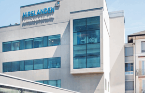
 Geneva, Switzerland Hirslanden Clinique La Colline
 -
 Geneva, Switzerland Generale-Beaulieu
 -
 Duisburg, Germany Duisburg-Nord Hospital
 -
 Istanbul, Turkey Acibadem Altunizade
 -
 Antalya, Turkey Hospital Medical Park Antalya
 -
 Dubai, UAE NMC Healthcare
 -
 Istanbul, Turkey Hospital “Memorial Şişli”
 -
 Milan, Italy San Raffaele University Hospital
 -
 Abu Dhabi, UAE Burjeel Hospital Abu Dhabi
 -
 Vienna, Austria Debling Private Clinic
 -
 Heidelberg, Germany Heidelberg University Hospital
 -
 Hamburg, Germany Asklepios Nord Heidberg
 -
 Winterthur, Switzerland Clinic "Lindberg"
 -
 Incheon, South Korea Gil Medical Center at Gachon University
 -
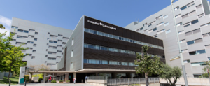
 Barcelona, Spain QuironSalud Barcelona Hospital
 -
 Barcelona, Spain Medical Center "Teknon"
 -
 Barcelona, Spain Sant Joan de Deu Children's Hospital
 -
 Barcelona, Spain University Hospital Barnaclinic+
 -
 Madrid, Spain University Clinic HM Madrid
 -
 Madrid, Spain University Hospital HM Monteprincipe
 -
 Zurich, Switzerland Hirslanden Clinic
 -
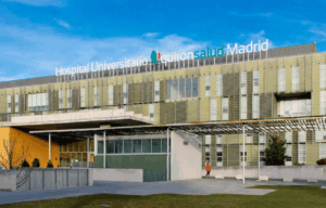
 Madrid, Spain Quiron Salud University Hospital
 -
 Duesseldorf, Germany Oncological Center Dusseldorf
 -
 Petah Tikva, Israel Schneider Children's Medical Center
 -
 Seoul, South Korea Samsung Medical Center
 -
 Seoul, South Korea SNUH


