


Fibroadenoma of the mammary gland
Disease Description
A benign neoplasm most often occurring in women under the age of 35. Fibroadenoma is a rounded, dense structure not adhered to the surrounding tissues, and in 85% of cases affects only one mammary gland.
According to WHO statistics, fibroadenoma is diagnosed in approximately 30% of women and rarely undergoes malignant transformation (develops into cancer).
Symptoms indicating the need for diagnosis and treatment
The disease may be asymptomatic and be discovered accidentally during self-examination or preventive screening.
Depending on the type of fibroadenoma, women may present with:
- Painful lump in the breast
- Discomfort in the mammary gland, increasing toward the end of the menstrual cycle
- Swelling and engorgement of the breast
- Rapid growth of the mass
- Increased nipple sensitivity
- Appearance of capillaries on the breast
Diagnostic and treatment methods
Diagnosis
An important diagnostic criterion is examination on the 5th-7th day of the menstrual cycle. It includes visual assessment of shape, symmetry, skin and nipple condition, as well as palpation of the mammary glands in various positions.
At the next stage, doctors in European clinics use instrumental methods:
- Ultrasound
- Mammography (X-ray of the mammary glands)
- MRI
- Biopsy
Treatment
The main method of treatment is surgical. For this, advanced tumor removal techniques are used:
- Enucleation (shelling) — without affecting the surrounding tissues
- Sectoral resection (for large neoplasms)
- Radio wave (at early stages)
Since mastopathy is associated with a disruption of hormonal rhythm, treatment will include hormone therapy for the prevention of relapses. Western medical centers use ultra-modern new-generation drugs that have passed all clinical trials and are distinguished by high therapeutic activity.
Innovations of world clinics
As a simultaneous diagnostic and treatment method, innovative vacuum aspiration biopsy under ultrasound control is used. Through mini-punctures with a diameter of 2 mm, trained specialists remove neoplasms up to 2—3 cm. The main advantage is the complete removal of the tumor while preserving the milk ducts and achieving an excellent cosmetic effect (without scars and hematomas). Hospitalization is not required for the woman to undergo the procedure, and within 2 hours she can return to her usual lifestyle.
Top clinics
-
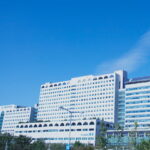
 Seoul, South Korea
Asan Medical Center
Seoul, South Korea
Asan Medical Center
-
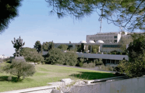
 Jerusalem, Israel
Hadassah Medical Center
Jerusalem, Israel
Hadassah Medical Center
-
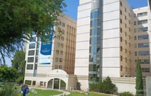 Petah Tikva, Israel
Medical Center Rabin
Petah Tikva, Israel
Medical Center Rabin
-

 Istanbul, Turkey
Medipol Mega University Hospital
Istanbul, Turkey
Medipol Mega University Hospital
-
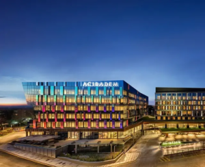 Istanbul, Turkey
Acibadem Altunizade
Istanbul, Turkey
Acibadem Altunizade
-
 Istanbul, Turkey
Acıbadem Ataşehir Clinic
Istanbul, Turkey
Acıbadem Ataşehir Clinic
-
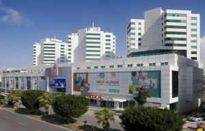 Antalya, Turkey
Hospital Medical Park Antalya
Antalya, Turkey
Hospital Medical Park Antalya
-
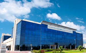
 Dubai, UAE
NMC Healthcare
Dubai, UAE
NMC Healthcare
-
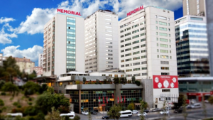 Istanbul, Turkey
Hospital Memorial Şişli
Istanbul, Turkey
Hospital Memorial Şişli
-
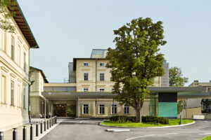
 Graz, Austria
Leech Private Hospital
Graz, Austria
Leech Private Hospital
-
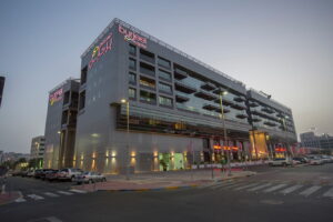 Abu Dhabi, UAE
Burjeel Hospital Abu Dhabi
Abu Dhabi, UAE
Burjeel Hospital Abu Dhabi
-
 Vienna, Austria
Debling Private Clinic
Vienna, Austria
Debling Private Clinic
-
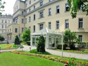 Vienna, Austria
Confraternität Private Hospital
Vienna, Austria
Confraternität Private Hospital
-

 Heidelberg, Germany
Heidelberg University Hospital
Heidelberg, Germany
Heidelberg University Hospital
-
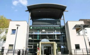 Hamburg, Germany
Asklepios Nord Heidberg
Hamburg, Germany
Asklepios Nord Heidberg
-
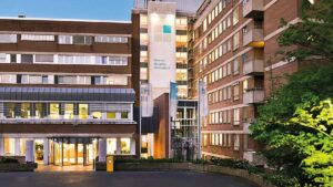 Dusseldorf, Germany
FKKD Clinical Complex
Dusseldorf, Germany
FKKD Clinical Complex
-
 Istanbul, Turkey
Memorial Bahçelievler Clinic
Istanbul, Turkey
Memorial Bahçelievler Clinic
-
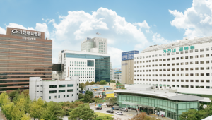 Incheon, South Korea
Gil Medical Center at Gachon University
Incheon, South Korea
Gil Medical Center at Gachon University
-
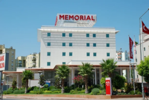 Antalya, Turkey
Memorial Antalya Hastanesi
Antalya, Turkey
Memorial Antalya Hastanesi
-
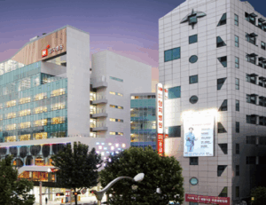 Seoul, South Korea
H+ Yangji Hospital
Seoul, South Korea
H+ Yangji Hospital
-
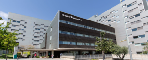
 Barcelona, Spain
QuironSalud Barcelona Hospital
Barcelona, Spain
QuironSalud Barcelona Hospital
-
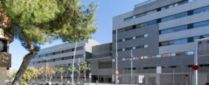 Barcelona, Spain
Dexeus University Hospital
Barcelona, Spain
Dexeus University Hospital
-
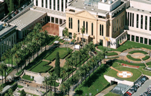 Barcelona, Spain
Medical Center Teknon
Barcelona, Spain
Medical Center Teknon
-
 Barcelona, Spain
University Hospital Barnaclinic+
Barcelona, Spain
University Hospital Barnaclinic+
-
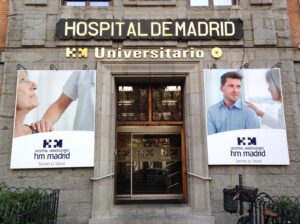 Madrid, Spain
University Clinic HM Madrid
Madrid, Spain
University Clinic HM Madrid
-
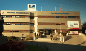 Madrid, Spain
University Hospital HM Monteprincipe
Madrid, Spain
University Hospital HM Monteprincipe
-
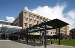 Hamburg, Germany
Asklepios Klinik Barmbek
Hamburg, Germany
Asklepios Klinik Barmbek
-
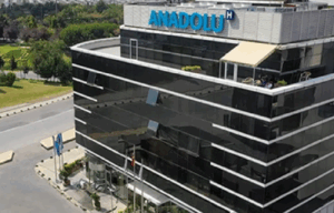 Gebze, Turkey
Anadolu Clinic
Gebze, Turkey
Anadolu Clinic
-
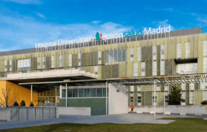 Madrid, Spain
QuironSalud University Hospital
Madrid, Spain
QuironSalud University Hospital
-
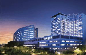 Seoul, South Korea
Samsung Medical Center
Seoul, South Korea
Samsung Medical Center
-
 Seoul, South Korea
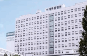
Medical Center at Ewha Womans University
Seoul, South Korea
Medical Center at Ewha Womans University
-
 Seoul, South Korea
SNUH
Seoul, South Korea
SNUH