


Nephroblastoma (Wilms Tumor)
Disease description
Nephroblastoma, also known as Wilms tumor, is a rare, highly malignant embryonal kidney tumor that predominantly occurs in children, most commonly between the ages of 2 and 5. This type of cancer is named after the German surgeon Max Wilms, who first described the disease in the early 20th century. Nephroblastoma accounts for approximately 5-7% of all pediatric malignancies. Despite its aggressive nature, it generally responds well to treatment, especially when diagnosed at an early stage.
Nephroblastoma primarily affects the kidneys — vital organs responsible for blood filtration and excretion of metabolic waste. The tumor can develop in one or both kidneys, although in most cases only one kidney is involved. Depending on the stage of the disease, the tumor may metastasize, that is, spread to other organs such as the lungs, liver, and lymph nodes.
Wilms tumor typically has a favorable prognosis, especially if the malignancy has not spread beyond the kidney. Remission is achieved in approximately 90% of pediatric patients
Symptoms
The tumor usually does not cause pain, and as a result, it often reaches a significant size by the time it is diagnosed. Most commonly, nephroblastoma is detected due to abdominal enlargement or the discovery of a painless, firm mass during palpation of the abdomen. In approximately 25% of cases, the diagnosis is made based on the presence of gross hematuria — visible blood in the urine.
Some symptoms may mimic gastrointestinal disorders and include abdominal pain, nausea, loss of appetite, and constipation. Fever, elevated blood pressure, and general malaise may also be observed. In rare cases, the tumor is identified due to the onset of pain.
Nephroblastoma is frequently diagnosed before metastases have developed, even when the primary tumor has reached a large size. This contributes to a relatively favorable prognosis, even in the presence of an aggressive (anaplastic) histologic subtype.
Diagnosis and treatment of Wilms Tumor in children
Diagnostics
Various imaging methods are used to assess the extent of nephroblastoma and to identify possible metastases. The primary diagnostic methods include abdominal and retroperitoneal ultrasound, as well as computed tomography (CT) and magnetic resonance imaging (MRI). A crucial step in diagnosis is a chest X-ray, as the lungs are the most common site of metastases in nephroblastoma.
To assess renal function and the overall condition of the patient, a 24-hour urine analysis and blood biochemistry tests are performed. The presence of erythrocytes in the urine (hematuria) may indicate renal involvement. Severe hematuria may lead to anemia, which can be confirmed by a complete blood count. To rule out the diagnosis of neuroblastoma, a urine test for catecholamines is also conducted.
For more detailed evaluation of the kidneys, excretory urography may be employed — an X-ray-based technique that helps assess renal anatomy and function. In the preoperative period, renal angiography with contrast agent may be recommended to visualize renal blood supply in greater detail.
A biopsy of the tumor is rarely performed before surgical intervention in cases of nephroblastoma, as disrupting the tumor capsule may contribute to dissemination. Diagnosis is typically based on imaging data.
Children with congenital anomalies that increase the risk of developing nephroblastoma should undergo regular kidney ultrasound screening for early detection of the tumor.
Treatment
Treatment of nephroblastoma typically involves a combination of surgery, chemotherapy, and radiotherapy. Given the rapid growth of Wilms tumor, therapy must begin immediately after diagnosis. The treatment strategy depends on the stage of the disease.
Chemotherapy may be involved prior to surgery in order to reduce the size of the tumor and facilitate its removal. This is especially important in stage III-IV cases, when the tumor is too large and its removal poses a risk of damaging vital structures. Preoperative chemotherapy also reduces the risk of tumor capsule rupture during surgery.
Following surgical resection, chemotherapy is routinely prescribed, and in some cases, radiotherapy is also involved to eliminate residual malignant cells.
In most cases, complete surgical removal of the affected kidney is performed (nephrectomy), sometimes including surrounding tissues, the adrenal gland, and regional lymph nodes (radical nephrectomy). In cases where both kidneys are involved or the patient has only one kidney, nephron-sparing surgery (partial nephrectomy) may be performed.
Innovations in global clinics
In rare cases, where the established treatment protocol does not yield the expected outcomes, high-dose chemotherapy followed by hematopoietic stem cell transplantation may be considered. This decision is made exclusively by a multidisciplinary team of highly qualified specialists in leading clinics in Europe and worldwide.
Procedures
Top clinics
-

 Baden-Baden, Germany
Max Grundig Clinic
Baden-Baden, Germany
Max Grundig Clinic
-

 Seoul, South Korea
Asan Medical Center
Seoul, South Korea
Asan Medical Center
-

 Jerusalem, Israel
Hadassah Medical Center
Jerusalem, Israel
Hadassah Medical Center
-

 Geneva, Switzerland
Generale-Beaulieu
Geneva, Switzerland
Generale-Beaulieu
-

 Istanbul, Turkey
Acibadem Altunizade
Istanbul, Turkey
Acibadem Altunizade
-
 Istanbul, Turkey
Medistate International Hospital
Istanbul, Turkey
Medistate International Hospital
-
 Antalya, Turkey
Hospital Medical Park Antalya
Antalya, Turkey
Hospital Medical Park Antalya
-

 Dubai, UAE
NMC Healthcare
Dubai, UAE
NMC Healthcare
-
 Istanbul, Turkey
Hospital Memorial Şişli
Istanbul, Turkey
Hospital Memorial Şişli
-

 Milan, Italy
San Raffaele University Hospital
Milan, Italy
San Raffaele University Hospital
-
 Abu Dhabi, UAE
Burjeel Hospital Abu Dhabi
Abu Dhabi, UAE
Burjeel Hospital Abu Dhabi
-

 Vienna, Austria
Debling Private Clinic
Vienna, Austria
Debling Private Clinic
-
 Heidelberg, Germany
Heidelberg University Hospital
Heidelberg, Germany
Heidelberg University Hospital
-
 Hamburg, Germany
Asklepios Nord Heidberg
Hamburg, Germany
Asklepios Nord Heidberg
-
 Dusseldorf, Germany
FKKD Clinical Complex
Dusseldorf, Germany
FKKD Clinical Complex
-
 Nyon, Switzerland
Clinique Genolier
Nyon, Switzerland
Clinique Genolier
-
 Istanbul, Turkey
Memorial Ataşehir Clinic
Istanbul, Turkey
Memorial Ataşehir Clinic
-
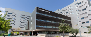
 Barcelona, Spain
QuironSalud Barcelona Hospital
Barcelona, Spain
QuironSalud Barcelona Hospital
-
 Barcelona, Spain
Medical Center Teknon
Barcelona, Spain
Medical Center Teknon
-
 Barcelona, Spain
Sant Joan de Deu Children's Hospital
Barcelona, Spain
Sant Joan de Deu Children's Hospital
-
 Barcelona, Spain
University Hospital Barnaclinic+
Barcelona, Spain
University Hospital Barnaclinic+
-
 Madrid, Spain
University Clinic HM Madrid
Madrid, Spain
University Clinic HM Madrid
-
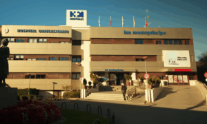 Madrid, Spain
University Hospital HM Monteprincipe
Madrid, Spain
University Hospital HM Monteprincipe
-
 Hamburg, Germany
Asklepios Klinik Barmbek
Hamburg, Germany
Asklepios Klinik Barmbek
-
 Gebze, Turkey
Anadolu Clinic
Gebze, Turkey
Anadolu Clinic
-
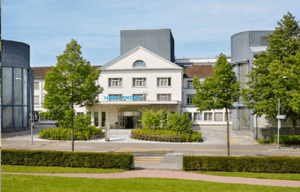 Zurich, Switzerland
Hirslanden Clinic
Zurich, Switzerland
Hirslanden Clinic
-
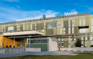 Madrid, Spain
QuironSalud University Hospital
Madrid, Spain
QuironSalud University Hospital
-
 Duesseldorf, Germany
Oncological Center Dusseldorf
Duesseldorf, Germany
Oncological Center Dusseldorf
-
 Petah Tikva, Israel
Schneider Children's Medical Center
Petah Tikva, Israel
Schneider Children's Medical Center
-
 Seoul, South Korea
Samsung Medical Center
Seoul, South Korea
Samsung Medical Center
-
 Seoul, South Korea
SNUH
Seoul, South Korea
SNUH